
MRI Scan in Chennai for Spine Injury – Advanced Imaging for Accurate Diagnosis
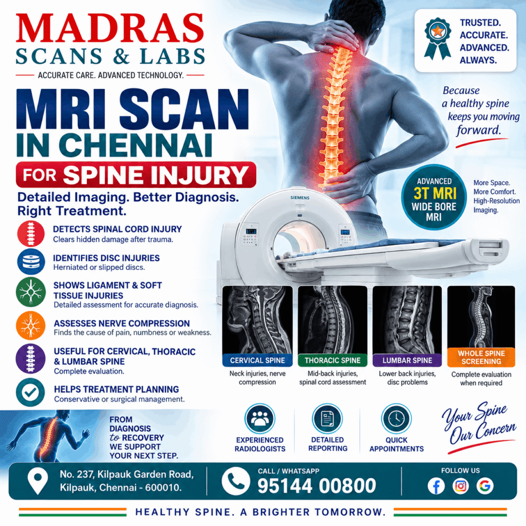
Spine injuries can occur due to road traffic accidents, falls, sports injuries, workplace accidents or sudden twisting movements. These injuries may affect the vertebrae, intervertebral discs, spinal cord, ligaments and surrounding soft tissues.
While some spine injuries cause only temporary discomfort, others can result in persistent pain, numbness, weakness or neurological complications.
An MRI Scan in Chennai can help doctors evaluate the spinal cord, nerves, discs and soft tissues in detail, identify the extent of an injury and determine the appropriate treatment.
At Madras Scans & Labs, Kilpauk, we offer advanced 3T MRI imaging for evaluating cervical, thoracic and lumbar spine injuries using our Siemens 3T Wide Bore MRI scanner.
Our MRI services help orthopaedic surgeons, neurologists and neurosurgeons assess spinal conditions and plan appropriate medical or surgical management.
1. Why Is an MRI Scan Required After a Spine Injury?
The spine consists of vertebrae, intervertebral discs, ligaments, nerve roots and the spinal cord. Injuries affecting these structures may cause pain, reduced mobility or neurological symptoms.
Although X-rays and CT scans provide valuable information about bone injuries, they may not adequately demonstrate damage to the spinal cord, ligaments and other soft tissues.
MRI uses powerful magnetic fields and radio waves to generate detailed images of these structures without ionizing radiation.
An MRI Scan for Spine Injury may be recommended when a doctor suspects:
- Spinal cord injury or compression.
- Herniated or ruptured intervertebral discs.
- Ligament tears or soft tissue injuries.
- Nerve root compression.
- Spinal epidural hematoma.
- Persistent neurological symptoms following trauma.
- Injury to the spinal cord despite inconclusive X-ray or CT findings.
MRI findings help doctors understand the nature and extent of an injury and determine whether conservative treatment, further investigation or surgical intervention is required.
Important: Following significant spinal trauma, CT is often the initial imaging examination for suspected vertebral fractures. MRI is used when additional evaluation of the spinal cord, ligaments, discs or neurological structures is clinically indicated.
2. What Types of Spine Injuries Can MRI Detect?
A. Spinal Cord Injury
The spinal cord carries nerve signals between the brain and the rest of the body. Damage to the spinal cord may result in weakness, numbness, sensory disturbances or paralysis.
MRI can demonstrate abnormalities such as spinal cord swelling, hemorrhage, compression and changes in spinal cord signal.
These findings help specialists assess the injury and plan further management.
B. Herniated or Slipped Disc
Intervertebral discs act as cushions between the vertebrae. Following trauma or degenerative changes, a disc may bulge or herniate and compress nearby nerve roots.
MRI can identify the location and extent of disc herniation and its relationship to the spinal cord or nerve roots.
Common symptoms include back pain, radiating leg pain, numbness and muscle weakness.
C. Spinal Ligament Injury
Ligaments stabilize the spine and maintain alignment between the vertebrae.
High-impact trauma may damage these ligaments, potentially affecting spinal stability.
MRI provides detailed visualization of spinal ligaments and surrounding soft tissues, helping specialists identify injuries that may not be apparent on conventional X-rays.
D. Nerve Root Compression
Spinal injuries can result in nerve compression due to disc herniation, displaced bone fragments or swelling of surrounding tissues.
MRI can help identify the location and possible cause of nerve compression.
Symptoms may include pain radiating into the arms or legs, tingling, numbness or weakness.
E. Spinal Epidural Hematoma
An epidural hematoma is a collection of blood within the spinal canal, outside the spinal cord.
It may develop following trauma and can compress neural structures.
MRI can help identify the location and extent of the hematoma.
Sudden weakness, difficulty walking or loss of bladder or bowel control following a spine injury requires immediate emergency medical evaluation.
3. MRI Cervical Spine Scan in Chennai – Evaluation of Neck Injuries
The cervical spine consists of seven vertebrae located in the neck.
Cervical spine injuries may occur following road accidents, falls, sports injuries or sudden acceleration and deceleration movements.
An MRI Cervical Spine Scan in Chennai may be recommended when a doctor suspects injury involving the cervical spinal cord, intervertebral discs, ligaments or nerve roots.
MRI can help evaluate:
- Cervical disc herniation.
- Spinal cord compression.
- Cervical ligament injuries.
- Nerve root compression.
- Spinal cord edema or hemorrhage.
- Soft tissue abnormalities following trauma.
Patients experiencing persistent neck pain, arm weakness, numbness or neurological symptoms following an injury may require further evaluation.
Do not move a person with a suspected serious neck or spinal injury unless necessary for immediate safety. Emergency medical assistance should be obtained.
4. MRI Thoracic Spine Scan in Chennai – Assessment of Mid-Back Injuries
The thoracic spine consists of twelve vertebrae and forms the middle portion of the spinal column.
Thoracic spine injuries may occur following high-impact trauma, falls or accidents.
MRI may be useful for evaluating spinal cord compression, ligament damage, disc injuries and other soft tissue abnormalities.
An MRI Thoracic Spine Scan can help identify:
- Thoracic disc herniation.
- Spinal cord compression.
- Ligament injuries.
- Epidural hematoma.
- Spinal cord abnormalities associated with trauma.
The choice of imaging depends on the clinical findings and the nature of the injury.
5. MRI Lumbar Spine Scan in Chennai – Evaluation of Lower Back Injuries
The lumbar spine consists of five vertebrae located in the lower back.
Lower back injuries can occur due to falls, lifting injuries, sports activities or road traffic accidents.
An MRI Lumbar Spine Scan in Chennai provides detailed images of the lumbar discs, spinal canal, nerve roots and surrounding soft tissues.
MRI may be recommended to investigate:
- Lumbar disc herniation.
- Nerve root compression.
- Traumatic ligament injuries.
- Spinal canal abnormalities.
- Persistent neurological symptoms following an injury.
Lumbar MRI can also help identify the cause of radiating leg pain when nerve compression is suspected.
6. MRI Whole Spine Screening in Chennai – When Is It Required?
In selected patients, doctors may recommend MRI imaging of more than one region of the spine.
An MRI Whole Spine Screening in Chennai may be useful when symptoms suggest abnormalities involving multiple spinal levels or when the clinical examination does not clearly identify the affected region.
Depending on the clinical indication, whole spine MRI can help evaluate:
- Spinal cord abnormalities at multiple levels.
- Multilevel disc disease.
- Spinal canal narrowing.
- Multiple areas of nerve compression.
- Other abnormalities involving different regions of the spine.
However, a whole spine screening examination is not a substitute for a dedicated MRI study of a specific region when detailed evaluation is required.
The appropriate MRI protocol should be selected according to the patient’s symptoms, examination findings and the referring doctor’s clinical requirements.
7. MRI Scan vs CT Scan for Spine Injury – Which Is Required?
Both MRI and CT play important roles in evaluating spine injuries. However, they provide different types of diagnostic information.
| Feature | MRI Scan | CT Scan |
|---|---|---|
| Spinal cord assessment | Detailed evaluation | Limited soft tissue detail |
| Vertebral fractures | Can demonstrate fractures and bone marrow changes | Excellent evaluation of bone fractures |
| Intervertebral discs | Detailed visualization | Limited compared with MRI |
| Spinal ligament injuries | Detailed evaluation | Limited direct visualization |
| Nerve root compression | Detailed evaluation | Less detailed soft tissue assessment |
| Radiation exposure | No ionizing radiation | Uses ionizing radiation |
| Examination duration | Generally longer | Generally faster |
| Emergency trauma assessment | Used for selected clinical indications | Often the initial examination for suspected bony injury |
CT and MRI are complementary investigations. The choice depends on the suspected injury, neurological findings, patient stability and the information required for treatment planning.
8. Advantages of 3T MRI for Spine Imaging
At Madras Scans & Labs, we provide spine imaging using advanced Siemens 3T Wide Bore MRI technology.
A 3 Tesla MRI scanner uses a strong magnetic field that can provide high-resolution images of the spinal cord, discs, ligaments and surrounding soft tissues.
Benefits of 3T MRI Technology
High-Resolution Imaging
Advanced MRI technology enables detailed visualization of spinal anatomy, helping radiologists identify abnormalities involving the spinal cord, discs and surrounding structures.
Detailed Soft Tissue Assessment
MRI provides excellent soft tissue contrast, allowing evaluation of spinal nerves, intervertebral discs, ligaments and other structures.
Wide Bore Design
Our Siemens Wide Bore MRI scanner is designed to provide a more spacious scanning environment, which may improve patient comfort.
No Ionizing Radiation
MRI does not use ionizing radiation, making it suitable for repeated examinations when clinically required.
Advanced Imaging Protocols
Dedicated spine MRI protocols can be selected according to the anatomical region and the referring doctor’s clinical requirements.
The suitability of 3T MRI depends on the patient’s condition, implanted medical devices and MRI safety assessment.
9. Who Should Undergo an MRI Scan After a Spine Injury?
Your doctor may recommend an MRI Scan in Chennai following a spine injury if you experience:
- Persistent neck or back pain associated with neurological symptoms.
- Numbness or tingling in the arms or legs.
- Weakness in the upper or lower limbs.
- Pain radiating from the spine into the extremities.
- Difficulty walking following an injury.
- Suspected spinal cord or ligament injury.
- Persistent neurological symptoms despite inconclusive initial imaging.
Not every patient with back pain or a minor spine injury requires MRI. The decision should be based on clinical examination and the suspected underlying condition.
When Is Emergency Medical Attention Required?
Seek immediate emergency care if a spine injury is accompanied by:
- New or progressive weakness in the arms or legs.
- Loss of sensation.
- Loss of bladder or bowel control.
- Severe neck or back pain following significant trauma.
- Difficulty walking or inability to move a limb.
These symptoms may indicate a serious spinal or neurological injury requiring urgent assessment.
10. How Is an MRI Spine Scan Performed?
An MRI spine examination is a non-invasive imaging procedure.
Step 1: Patient Preparation
The radiology team reviews the patient’s medical history and checks for implanted medical devices, metallic foreign bodies and other MRI safety considerations.
Step 2: Positioning
The patient is positioned on the MRI examination table. Appropriate support and positioning devices may be used to improve comfort.
Step 3: MRI Imaging
The scanner obtains images of the required spinal region using dedicated imaging sequences.
Patients are generally asked to remain still during image acquisition to minimize motion artifacts.
Step 4: Image Interpretation
The acquired images are reviewed by a radiologist, who prepares a diagnostic report.
Step 5: Clinical Consultation
The patient should discuss the MRI findings with the referring doctor, who will determine the appropriate treatment based on the imaging results and clinical examination.
How Long Does an MRI Spine Scan Take?
A dedicated MRI examination of one spinal region commonly takes approximately 20–40 minutes.
The duration may vary depending on the clinical indication, imaging protocol and whether additional sequences or contrast administration are required.
Whole spine examinations may take longer.
11. Why Choose Madras Scans & Labs for MRI Scan in Chennai?
Madras Scans & Labs provides advanced diagnostic imaging services for patients requiring MRI examinations of the brain, spine, joints and other anatomical regions.
Our MRI imaging services include:
- Siemens 3T Wide Bore MRI technology.
- High-resolution imaging of the cervical, thoracic and lumbar spine.
- MRI Whole Spine Screening.
- Detailed assessment of intervertebral discs and spinal soft tissues.
- Experienced radiology reporting.
- Convenient appointment scheduling.
- Accessible location in Kilpauk, Chennai.
Our team focuses on providing quality diagnostic imaging and a comfortable patient experience.
12. Book Your MRI Scan in Chennai at Madras Scans & Labs
If your doctor has recommended an MRI examination following a spine injury, contact Madras Scans & Labs for appointment information.
Our advanced 3T MRI technology supports detailed evaluation of spinal anatomy and assists your treating doctor in diagnosis and treatment planning.
MADRAS SCANS & LABS
📍 No. 237, Kilpauk Garden Road, Kilpauk, Chennai – 600010
📞 Call / WhatsApp: 95144 00800
🌐 Website: https://madrasscan.com
Advanced 3T MRI Scan in Chennai – Accurate Imaging for Better Clinical Decisions.