
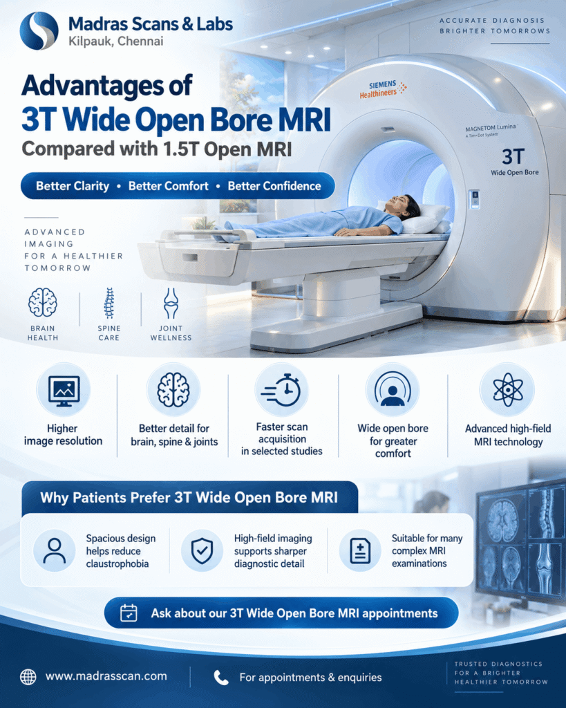
3T Wide Open Bore MRI vs 1.5T Open MRI: Understanding the Advantages
Experience advanced MRI technology with greater imaging detail and patient comfort
Magnetic Resonance Imaging (MRI) is one of the most important diagnostic tools in modern healthcare. It provides detailed images of the brain, spine, joints, muscles, nerves and internal organs without using ionising radiation.
However, not all MRI scanners offer the same imaging capabilities.
When patients search for an MRI Scan in Chennai, they frequently encounter terms such as 1.5T MRI, 3T MRI, Open MRI and Wide Open Bore MRI.
Understanding the differences can help patients select an appropriate scanning facility in consultation with their referring doctor.
At Madras Scans & Labs, Chennai, our Siemens 3T Wide Open Bore MRI system combines high magnetic field strength with a spacious scanner design, offering high-resolution diagnostic imaging while prioritising patient comfort.
1. What is the difference between 3T MRI and 1.5T MRI?
The letter T stands for Tesla, the unit used to measure the strength of an MRI scanner’s magnetic field.
A 3T MRI scanner operates at a magnetic field strength of 3 Tesla, whereas a 1.5T MRI operates at 1.5 Tesla.
This means that a 3T MRI has twice the magnetic field strength of a 1.5T MRI.
The increased field strength generally provides a higher signal-to-noise ratio (SNR), which can be used to obtain finer imaging detail, reduce acquisition times or balance both advantages.
Importantly, twice the magnetic strength does not automatically mean twice the image quality or twice the diagnostic accuracy. The final image quality also depends on scanner hardware, imaging protocols, patient movement and the examination being performed.
2. What is a 3T Wide Open Bore MRI scanner?


A 3T Wide Open Bore MRI scanner combines two important features:
High-field 3 Tesla imaging: Provides stronger MRI signals that can support detailed anatomical evaluation.
Wide-bore design: Offers a larger patient opening compared with traditional narrow-bore MRI systems.
The wider opening can help patients who experience anxiety in enclosed spaces and can improve positioning for some patients with larger body sizes.
It is important to understand that a wide open bore MRI is not the same as a completely open-sided MRI scanner. A wide-bore system still has a cylindrical opening, whereas a true open MRI has a different magnet configuration.
Both 1.5T and 3T scanners can be manufactured with wide-bore designs. Therefore, field strength and scanner openness should be considered separately.
3. Seven important advantages of 3T Wide Open Bore MRI
Advantage 1: Higher image resolution and anatomical detail
One of the main technical advantages of 3T MRI is its higher available signal-to-noise ratio.
This additional signal can be used to acquire thinner image slices or improve spatial resolution while maintaining clinically useful image quality.
Detailed imaging can be particularly valuable for examining smaller anatomical structures.
For example:
Brain MRI: Detailed evaluation of brain anatomy and selected neurological abnormalities.
Knee MRI: Evaluation of menisci, ligaments, cartilage and other joint structures.
Spine MRI: Visualisation of discs, spinal cord and nerve-related structures.
Small-joint MRI: Detailed assessment of selected wrist, ankle and other musculoskeletal abnormalities.
The degree of improvement depends on the clinical application and imaging technique.
Advantage 2: Potential for shorter scan acquisition times
Patients often ask, “How much time does a 3T MRI scan take?”
A 3T MRI scanner can use its increased signal to accelerate certain imaging sequences while maintaining adequate image quality.
This can reduce the duration of selected scan acquisitions compared with equivalent protocols on a 1.5T system.
However, the total examination time depends on the body part, number of imaging sequences, contrast requirements and patient cooperation.
A 3T MRI is not necessarily faster for every examination.
Advantage 3: Greater patient comfort with a wide-bore design
A conventional narrow-bore MRI may be uncomfortable for patients who experience claustrophobia.
A wide-bore MRI provides additional space around the patient and may make scanning more tolerable.
Potential benefits include:
A more spacious examination environment.
Improved accommodation for certain patients with larger body sizes.
Easier positioning for some examinations.
Less perceived confinement for some patients.
The experience varies between individuals. Patients with severe claustrophobia may still require additional support or a genuinely open-sided MRI system.
Advantage 4: Detailed brain and neurological imaging


3T MRI is particularly useful in several neurological imaging applications.
Higher signal strength can support detailed structural imaging and certain advanced MRI techniques, including susceptibility-weighted imaging and functional MRI where available.
A 3T MRI may be particularly helpful in appropriately selected examinations for:
Epilepsy evaluation.
Multiple sclerosis assessment.
Selected small structural brain abnormalities.
Detailed brain anatomy.
Certain vascular and advanced neurological examinations.
Whether 3T provides a meaningful diagnostic advantage depends on the suspected condition and the imaging protocol.
Advantage 5: Improved visualisation of small musculoskeletal structures


MRI is commonly used to evaluate joint injuries, spinal abnormalities and musculoskeletal conditions.
In selected examinations, the additional signal available at 3T can support high-resolution assessment of:
Knee ligaments and menisci.
Articular cartilage.
Shoulder tendons and related soft tissues.
Wrist and ankle structures.
Spinal discs and surrounding soft tissues.
This can be useful when the referring doctor requires detailed evaluation of small anatomical structures.
Nevertheless, a properly performed 1.5T MRI remains diagnostically effective for many common musculoskeletal conditions.
Advantage 6: Advanced imaging possibilities
High-field 3T MRI can offer technical advantages for specialised imaging applications.
Depending on scanner configuration, available software and the examination protocol, these may include:
Diffusion-weighted imaging: Used in brain imaging and selected other examinations.
Magnetic resonance angiography: Evaluation of blood vessels without necessarily requiring catheter angiography.
MR spectroscopy: Provides information about selected tissue metabolites.
Susceptibility-weighted imaging: Particularly useful for detecting magnetic susceptibility effects associated with blood products and certain other tissue characteristics.
Not every 3T scanner offers every advanced examination, and the availability of individual applications should be confirmed with the diagnostic centre.
Advantage 7: A balance between high-field imaging and a spacious scanner
The combination of 3T magnetic field strength and a wide-bore design provides two distinct benefits.
The high magnetic field can support detailed imaging, while the larger opening helps address some of the comfort concerns associated with traditional narrow MRI scanners.
For patients who require high-resolution imaging but are concerned about enclosed spaces, a 3T Wide Open Bore MRI may be a suitable option.
4. Comparison: 3T Wide Open Bore MRI vs 1.5T Open MRI
Feature | 3T Wide Open Bore MRI | 1.5T Open MRI |
|---|---|---|
Magnetic strength | 3 Tesla | 1.5 Tesla |
MRI signal | Generally higher available SNR | Lower available SNR under comparable conditions |
Image resolution | Can support finer anatomical detail | High-quality imaging possible |
Scan acquisition speed | Can be faster for selected protocols | Depends on hardware and protocol |
Patient opening | Wide cylindrical bore | Depends on whether genuinely open-sided or wide-bore |
Claustrophobia comfort | More spacious than narrow-bore systems | True open-sided designs may offer greater openness |
Brain imaging | Advantages in selected advanced applications | Suitable for many routine and specialised examinations |
Joint imaging | Can support high-resolution anatomical assessment | Effective for many routine joint examinations |
Implant compatibility | Requires device-specific 3T safety verification | Some devices are approved only under 1.5T conditions |
Overall suitability | Depends on the clinical question | Depends on the clinical question |
Important distinction: A 1.5T scanner described as open may refer either to a truly open-sided configuration or to a wide-bore cylindrical scanner. Confirm its actual design before comparing patient comfort.
5. Is a 3T MRI always better than a 1.5T MRI?
Not necessarily.
Although 3T MRI offers technical benefits, there are situations in which a 1.5T system may be preferred.
For instance, imaging near certain metallic implants can be more challenging at 3T because susceptibility-related artefacts may be more pronounced. Some implanted medical devices also have MRI-conditional labelling that permits scanning at 1.5T but not at 3T.
At 3T, radiofrequency energy deposition and certain image artefacts also require careful management.
The radiologist and referring physician should select the scanner and protocol based on the patient’s clinical needs and safety requirements.
6. Who may benefit from a 3T Wide Open Bore MRI scan?
Patients may benefit from considering a 3T wide-bore examination when they require:
Detailed brain or neurological imaging.
High-resolution knee, shoulder or other joint imaging.
Selected complex spine examinations.
Specialised MRI protocols where 3T offers an advantage.
A more spacious alternative to a conventional narrow-bore scanner.
For patients with significant claustrophobia, implanted medical devices or other individual requirements, the suitability of the scanner should be assessed before booking.
7. 3T Wide Open Bore MRI Scan in Chennai – Madras Scans & Labs

At Madras Scans & Labs, we understand that patients look for both diagnostic imaging quality and a comfortable scanning experience.
Our Siemens 3T Wide Open Bore MRI system offers high-field magnetic resonance imaging with a spacious scanner design.
Patients referred for brain MRI, spine MRI, joint MRI and other indicated MRI investigations can enquire about available scan protocols and appointment requirements.
Our team can also guide patients regarding examination preparation, MRI safety screening and the information required before undergoing a scan.
Why choose Madras Scans & Labs for your MRI examination?
Siemens 3T Wide Open Bore MRI technology.
High-field MRI imaging capability.
A spacious scanner design.
MRI examinations for multiple anatomical regions.
Patient preparation and safety screening before scanning.
Convenient diagnostic services in Kilpauk, Chennai.
MRI Scan in Chennai – Madras Scans & Labs
For appointments and MRI enquiries, visit www.madrasscan.com.
Frequently asked questions
1. Is 3T MRI better than 1.5T MRI?
3T MRI offers higher magnetic field strength and generally greater available signal, which can support finer anatomical detail or faster acquisition in selected examinations. However, a 1.5T MRI is sufficient for many diagnostic applications, and some examinations are better suited to 1.5T.
2. Is 3T MRI more comfortable than a 1.5T open MRI?
Comfort depends primarily on the scanner’s physical design rather than magnetic field strength. A 3T wide-bore scanner provides a larger opening than a traditional narrow-bore system, but a genuinely open-sided 1.5T MRI may feel more spacious to certain patients.
3. Does a 3T MRI scan take less time?
Some sequences can be acquired more quickly at 3T. The overall examination may not be shorter because scanning time depends on the protocol and clinical requirements.
4. Is 3T MRI safe?
A 3T MRI is widely used in clinical practice when appropriate safety protocols are followed. Patients must inform the MRI team about pacemakers, cochlear implants, aneurysm clips, neurostimulators, other implants and any possible metallic foreign bodies.
5. Does a 3T MRI use radiation?
No. MRI uses magnetic fields and radiofrequency pulses rather than ionising radiation.
6. Can patients with claustrophobia undergo a 3T Wide Open Bore MRI?
Many patients who find traditional narrow-bore scanners uncomfortable may tolerate a wide-bore scanner better. However, patients with significant claustrophobia should discuss their concerns with the scanning team before the appointment.
7. Where can I get a 3T Wide Open Bore MRI Scan in Chennai?
Patients looking for a 3T Wide Open Bore MRI Scan in Chennai can enquire with Madras Scans & Labs, Kilpauk, about the available examinations and appointment arrangements.
Conclusion
The principal advantage of a 3T Wide Open Bore MRI is its combination of high magnetic field strength and a relatively spacious scanning environment.
Compared with a 1.5T MRI, 3T technology provides additional signal that can be used to improve image resolution, accelerate selected scanning sequences or support specialised MRI applications.
However, the appropriate MRI system depends on the examination, patient characteristics, safety considerations and clinical question.
At Madras Scans & Labs, Chennai, our Siemens 3T Wide Open Bore MRI technology supports advanced diagnostic imaging with an emphasis on patient comfort and safety.
For MRI Scan appointments in Chennai, visit madrasscan.com.