- Home
- Departments
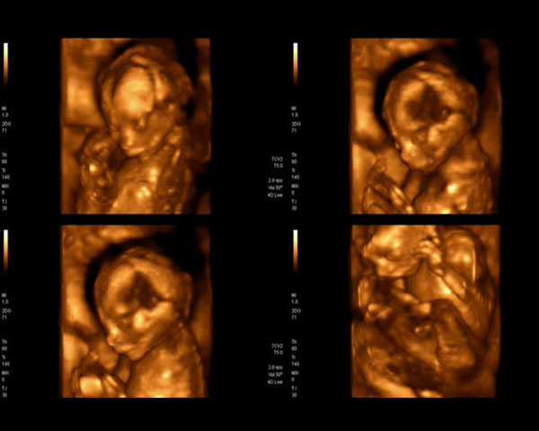
- 3D-4D Pregnancy Scan
3D/4D Pregnancy UltraSound Scan in Chennai
Ultrasounds in 3D and 4D are performed to examine closely suspected fetal anomalies, such as cleft lip and spinal cord issues, or to monitor something specific. keep in mind, 3D and 4D ultrasounds are usually not part of routine prenatal exams. There is a full range of ultrasound packages available for your entire pregnancy journey when you scan at Madras Scans.
on 100+ reviews.
When you scan with us, we use high-end ultrasound machines for every scan, along with the latest scanning technology experience of 4D scan as an option. a scan is the most common and reliable form of investigation in pregnancy. Extra dimensions provided through 3D, 4D scans give better pictures of your baby and more detailed information about your baby health. At Madras Scans, the aim of conducting a scan during pregnancy is to monitor you baby growth as well as to detect any abnormalities in organs structure, growth, position of placenta or amniotic fluid.
Madras Scans centre in chennai is a Centre for Excellence for Pregnancy Sonography and is the preferred referral centre for several leading Gynaecologists in Chennai.
We specialize in performing the following Ultrasound scans at different periods of gestation.
- Early Pregnancy Scan (7-11 Weeks)
- NT/NB Scan (11-13 Weeks)
- Combined First Trimester Screening (11-13 Weeks)
- Anomaly Scan (Level II Scan) (18 – 21 Weeks)
- Fetal Echocardiography (22 -24 Weeks)
- Well Being Scan (25 – 40 Weeks)
- 2D/ 3D /4D ultrasound scans are performed by experienced doctors.
Uses of the ultrasound
Ultrasound may be used at various points during pregnancy:
- First trimester - ultrasound performed within the first 3 months of pregnancy is used to check that the embryo is developing inside the womb (rather than inside a fallopian tube, for example), confirm the number of embryos, and calculate the gestational age and the baby’s due date.
- Second trimester - ultrasound performed between weeks 18 and 20 is used to check the development of fetal structures such as the spine, limbs, brain and internal organs. The size and location of the placenta is also checked. The baby’s sex can be established, if the parents wish to know.
- Third trimester - ultrasound performed after 30 weeks is used to check that the baby is continuing to grow at a normal rate. The location of the placenta is checked to make sure it isn’t blocking the cervix.

